 (1)
(1)Natusch MKH, Boothroyd CB, Botton GA, Humphreys CJ, and Krivanek OL *
Department of Materials Science and Metallurgy, University of
Cambridge, Pembroke Street, Cambridge CB2 3QZ, UK and
*Cavendish
Laboratory, Madingley Road, Cambridge CB3 0HE, UK
A theoretical model that predicts the detection limits of electron energy loss spectroscopy (EELS) would be very useful. It would allow one to check whether an element present at a suspected concentration should be detectable in an EELS spectrum acquired under particular conidtions. It would also show how to optimise acquisition and processing parameters so that the detection limits become as low as possible.
We are developing such a model based on synthesising EELS spectra corresponding to given concentrations of elements. The signal-to-noise ratios (SNRs) of spectral features due to elements of interest are then evaluated, and the detectability of the elements is judged using the Rose criterion (SNR > 4). Our current approach is focused on standardless Egerton-type quantification [1] involving the comparison of background-subtracted inner shell loss edges with theoretical cross-sections, and on minimum mass fraction (MMF) limits. In future work we plan to cover multiple least-square fitting of first and second difference spectra with standard spectra acquired from reference samples, and also minimum detectable mass (MDM) limits.
In order to be able to synthesise a spectrum, one must know accurately:
1) the total electron dose delivered to the sample per second,
2) the number of atoms of each atomic species per unit area (areal density), or the volume density of each species plus the specimen thickness,
3) the spectrum acquisition parameters (primary energy, illumination and collection angles, acquisition time),
4) the performance of the spectrometer and the detector ( detective quantum efficiency (DQE) of the detector, conversion efficiency (counts per fast electron), contribution to spectrum background, energy resolution),
5) the theoretical differential cross-sections for all the edges in front of the energy interval of interest for all the elements in the sample,
6) the contribution of plasmon and valence-type excitations to the high energy parts of the spectrum, and
7) all sample-modifying effects (contamination, radiation damage). For MDM type of detection limits, one must also know accurately the incident electron beam profile and how this spreads out in the specimen.
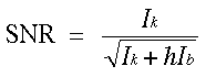
Once the model spectrum is synthesised, the detection limit is determined by computing the SNR of each edge of interest. The noise is evaluated according to Egerton's formula:
(1)
where Ik is the characteristic edge signal, Ib is the background integral and the dimensionless parameter h describes the variance of Ik due to fitting and extrapolation errors.
In our initial exploration of the problem, we have taken a number of simplifying assumptions. First, we have assumed that the detector has DQE = 1. We are primarily interested in the high counts regime necessary for exploring low concentrations. In this regime, the dominant DQE limit arises from channel-to-channel gain variations, and we are removing these using an algorithm that has been shown to reduce the variations by around 30x [2]. Second, we are modelling the plasmon and valence-loss contribution to the spectrum using Drude-Lorentz type oscillators [3], adjusted to match the observed plasmon energy and intensity. Third, we neglect contamination and radiation damage. The theoretical inner-shell cross-sections are computed using the Hartree-Slater routines provided by Gatan's EL/P software version 3.0.
We have applied the preliminary model to MgO crystals which showed that the plasmon and valence-loss contribution is overestimated in the model. If, however, the intensity in the model is adjusted to give the observed intensity in the experimental spectrum, preliminary predictions can be made. Under the assumption that this procedure is valid the model can be applied to the NIST K546 standard reference material [4], which contains Th and U at trace level concentrations of 0.010 atom % and 0.013 atom % respectively. Figure 1 shows an experimental spectrum of background-subtracted Th and U M4,5 edges. These were predicted as detectable by the present model with a Th M4,5 SNR of 9.1 and a U M4,5 SNR of 16.5, even though published spectra from the material have not revealed them to be present. Our model therefore correctly suggests that with optimised acquisition and processing parameters it should be possible to detect Th and U at trace level concentrations of 0.01 atom % in the NIST reference material.
We are grateful to Dr. D.E. Newbury for the NIST reference sample and to Gatan Inc. for financial support (MKHN).
1. Egerton RF (1986), Electron Energy-Loss Spectroscopy in the Electron Microscope, Plenum Press, New York
2. C.B. Boothroyd, K. Sato, K. Yamada (1990) 12th P Int C El Mic 2 : 80-81
3. Ritchie RH, Howie A (1977) Phil Mag 36: 463-481
4. Leapman RD, Newbury DE (1993) Anal Chem 65: 2409
FIGURE 1:
![[Figure 1]](fig1.jpg)
FIGURE 1: Experimental spectrum acquired at 100 kV with an incident beam dose of 9 uC compared to theoretical cross-sections of Th M4,5 and U M4,5 edges after background subtraction (Ib = 1.35x10⁸ counts).